
|
|
Lymphocytic thyroiditis - case 786
|
|


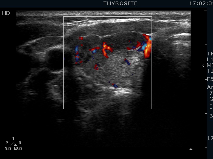
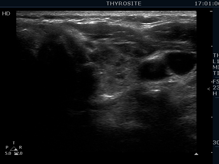
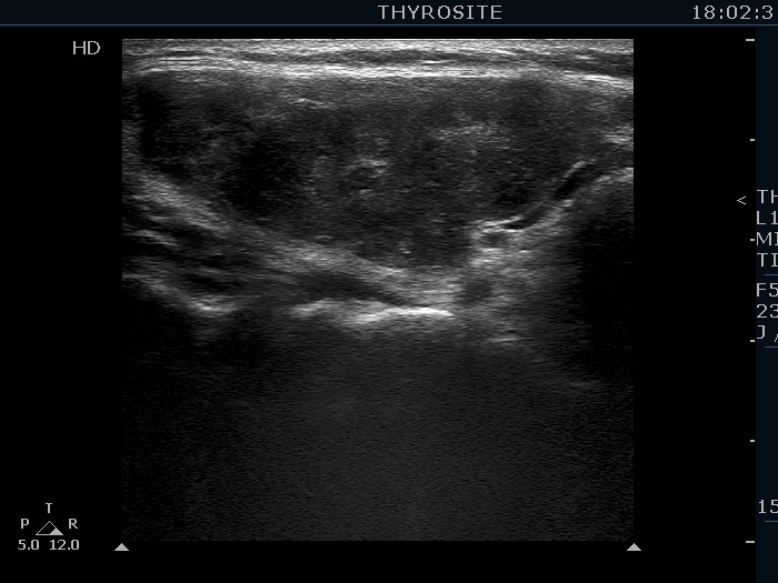

First examination (first row of images):
Clinical data: A 37-year-old woman requested an evaluation of complaints suggesting hypothyroidism. After her second birth two years ago, she was left with an excess weight of 18 kg.
Palpation: no abnormality.
Laboratory tests: TSH 3.52 mIU/L, FT4 14.1 pM/L, aTPO 405 U/mL.
Ultrasonography. The thyroid was echonormal and presented numerous hypoechogenic discrete areas. The echogenicity index was approximately 30%. None of the discrete areas corresponded to pathological nodule. The vascularity was a bit lower than the average.
Suggestion: TSH in a year.
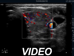
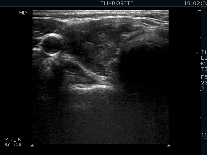
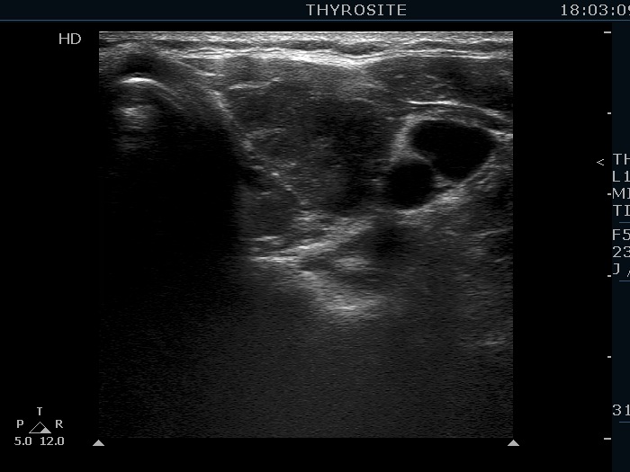
Second examination 19 months later (second row of images):
Comment. This is a typical presentation of a newly developed hypothyroidism regarding the change in echo pattern. Although the correlation between the echogenicity index and the hormonal status is week, in a particular patient, a developing hypothyroidism from an euthyroid state usually correlates with an increasing echogenicity index.Clinical data: This time the patient had no complaints, however the TSH was 16.8 mIU/L two month ago.
Palpation: no abnormality.
Laboratory tests: TSH 29.1 mIU/L, FT4 13.1 pM/L, aTPO 638 U/mL.
Ultrasonography. Compared with The previous examination, one change could be observed: the echogenicity index rose to 70-90%.
Daily 75 microgram levothyroxine was administered.