
|
|
Parathyroid lesions - case 976
|
|

Clinical data: A 63-year-old woman was referred for exact localization of a parathyroid before surgery. The patient had diffuse complaints, including fatigue, depression, weight loss. On routine laboratory blood test, an elevated serum calcium level, thereafter an elevated parathormone level (154 pg/mL) were detected. MIBI scintigraphy disclosed parathyroid adenoma corresponding to the right lower parathyroid.
Palpation: no abnormality.
Laboratory tests: TSH 1.17 mIU/L, parathormone 154 pg/ml, serum calcium 281 mM/L, phosphorus 0.99 mM/L. 24-hour urinary calcium content mM/L (normal value 2.5-7.5).
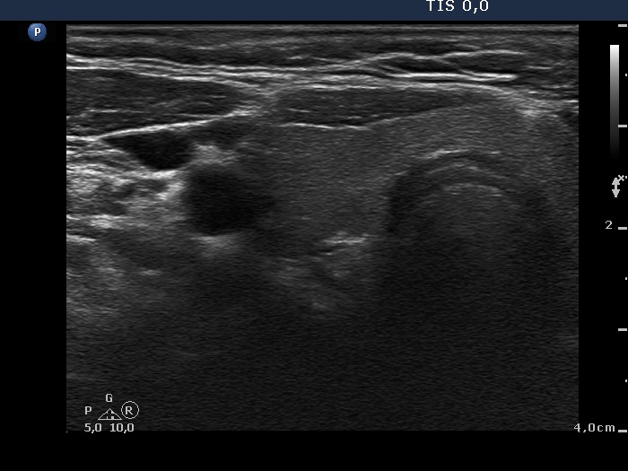
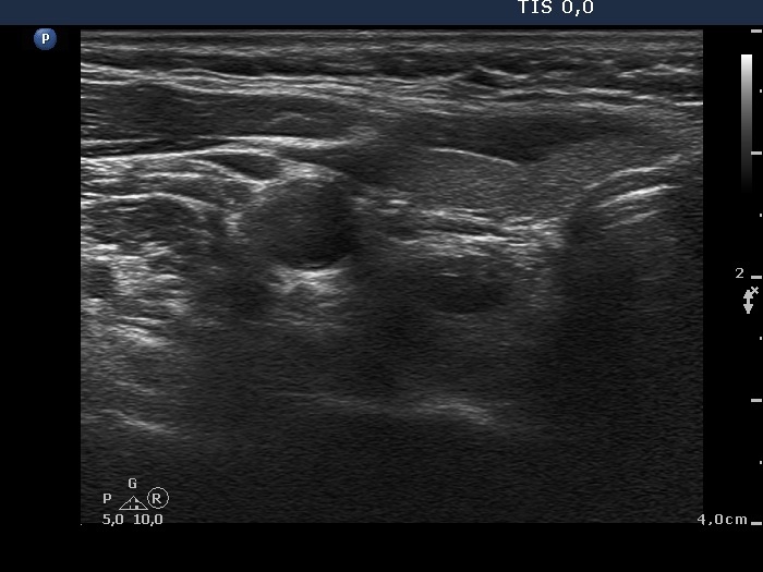
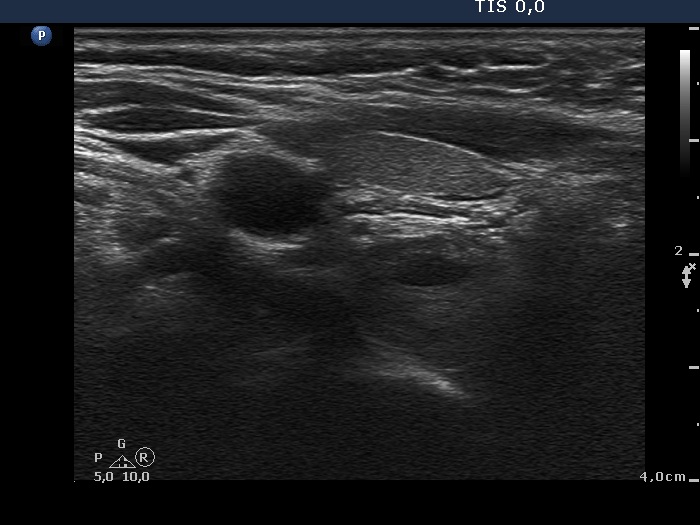
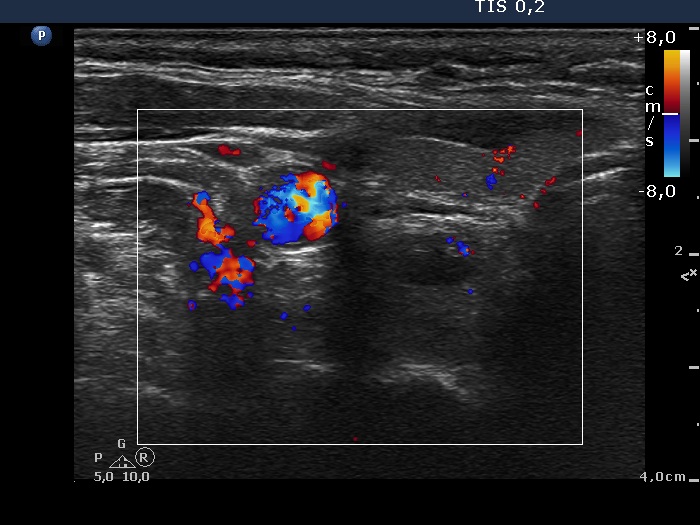
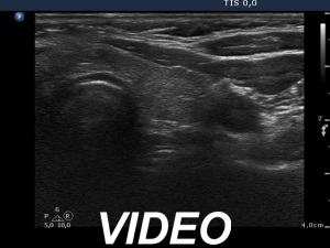
Ultrasonography. The thyroid was minimally hypoechoic and had several insignificant hypoechoic lesions. There was a solid-cystic mass dorsal to the lower pole of the right lobe.
Aspiration cytology of the lesion was performed. There were only scattered, non-atypical epithelial cells on the smear which was gained form a small amount of serous tissue.
Additional tests: Wash-out thyroglobulin resulted in 203 ng/mL, wash-out parathormone did in 134 pg/mL.
Histopathology: parathyroid adenoma.
Comment. The results of wash-out were not fully convincing in this case.