
|
|
Thyroid cancers - case 1041
|
|


Clinical data: A 51-year-old man was referred for evaluation of a nodular goiter which was discovered on lung screening which detected a substernally spreading right thyroid lobe. The patient told as that he has been aware of his goiter for more than 20 years. He had no clinical complaints.
Palpation: The right lobe was hugely enlarged and nodular on palpation.
Laboratory tests: TSH 0.11 mIU/L, FT4 14.6 pM/L.
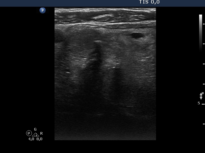
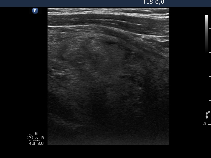
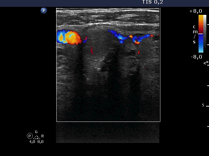
Ultrasonography. A large hypoechoic nodule occupied almost the entire right lobe. The lesion had both microcalcifications and coarse calcifications. The dimensions of the lobe were 58x55xminimum 80 mm, width, depth, length, respectively. The lower pole of the lobe did not come into sight while the patient swallowed. The nodule showed signs of perinodular blood flow. There was a hypoechoic nodule in the left lobe.
Aspiration cytology of the large nodule resulted in follicular tumor.
Before the operation, the surgeon reviewed the CT scan and three foci suspicious of metastasis were found in the right lung. During total thyroidectomy, thoracotomy had to be performed. Histopathology disclosed a widely invasive follicular cancer. Further examinations revealed that the suspicious lung lesions take up radioiodine.
Comments. This is a Grade 3 substernal spread. The distance between the most ventral and most dorsal part of the lowest visible section of the right lobe was measured to be 32 mm. It means a substantial degree of substernal spread.