
|
|
Thyroid cancers - case 649
|
|

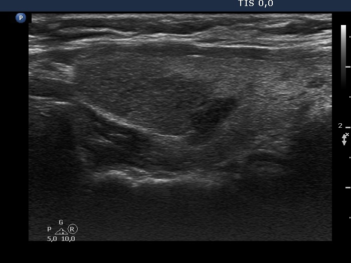
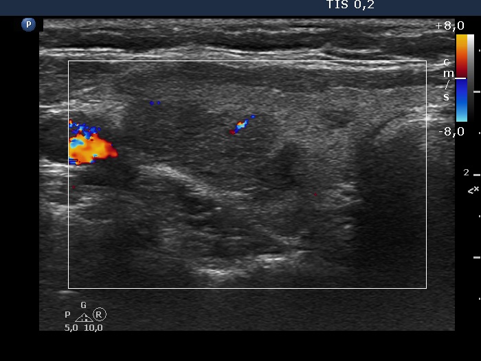
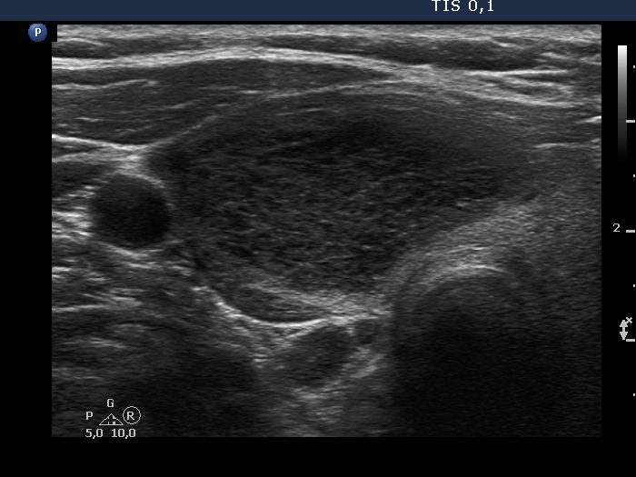
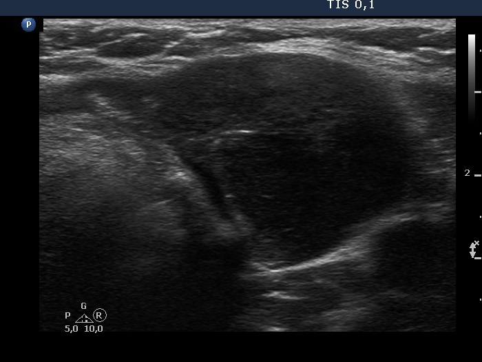
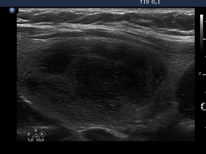
Right lobe


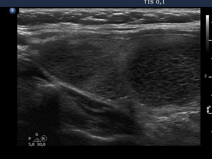
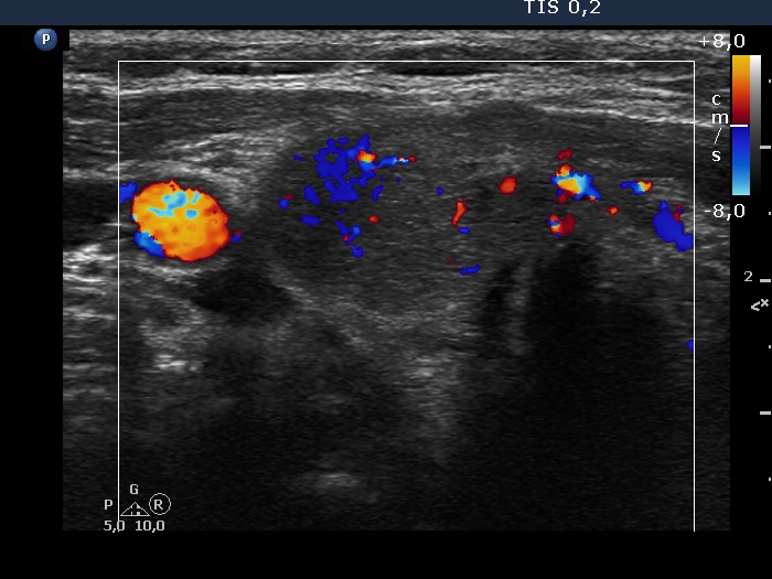
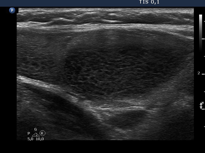
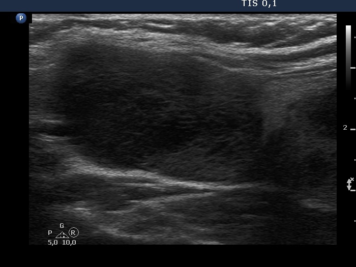
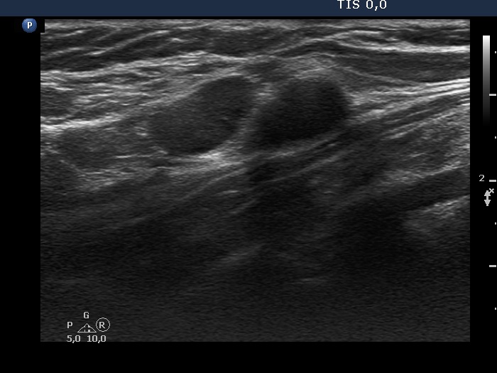
Left lobe
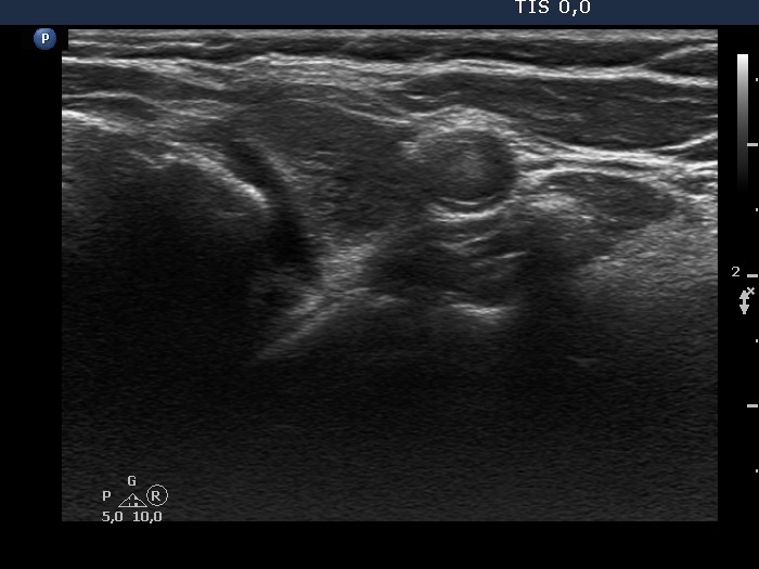

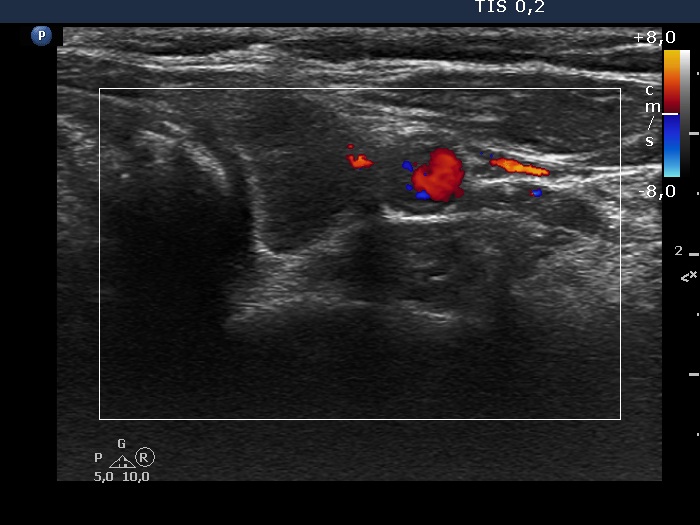


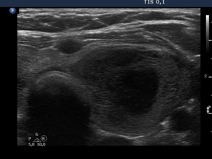
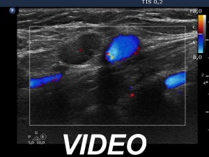
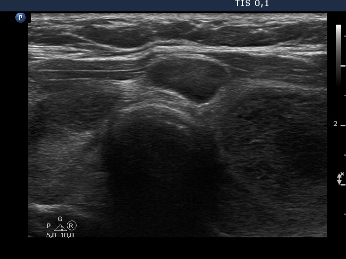
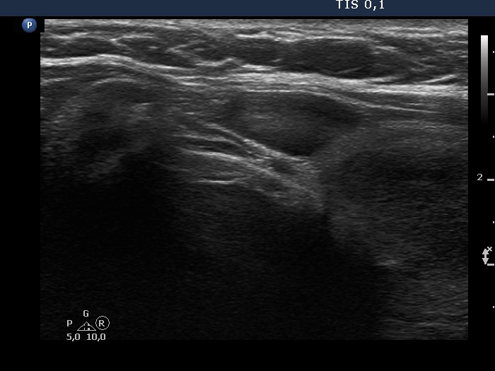
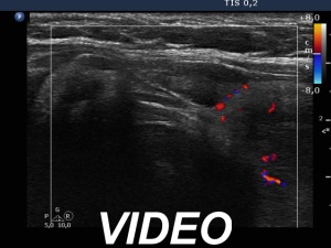
Neck lymph nodes

First examination (first, third and fifth rows of images):
Clinical presentation: A 48-year-old woman requested a thyroid evaluation. She was diagnosed with a low grade, stage III/A non Hodgkin lymphoma two years ago. On the first and subsequent PET CT scan, no progression was detected, and on all occasions multiple foci were described within the thyroid. The patient hasn't got any therapy yet.
Palpation: Both thyroid lobes were a bit firm, but no discrete lesions could be palpated. Numerous firm lymph nodes were palpable at both sides of the neck.
Laboratory tests: TSH 1.56 mIU/L, anti-TPO 0 U/mL, ahTg below 20 U/mL.
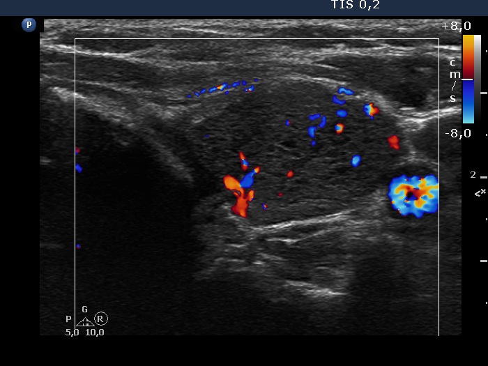
Ultrasonography. The thyroid was echonormal and contained multiple inhomogeneous, partly blurred hypoechogenic and moderately hypoechogenic discrete lesions. Multiple lymph nodes were found on both sides of the neck.
Cytology was performed from 3 different thyroid lesions and resulted in a pattern corresponding to non Hodgkin lymphoma.
Second examination 3 years later (second, fourth and sixth rows of images):
Clinical presentation: The patient requested a repeat examination because she has had already neck discomfort and occasionally difficulties in swallowing. She first noticed these complaints 3 months ago. She was under regular oncology check-up and was last checked for 2 months. At the time, it was still not considered warranted to treat lymphoma.
Palpation: Both thyroid lobes were enlarged and firm lesions were palpable in both lobes.
Laboratory tests: TSH 1.98 mIU/L, anti-TPO 4.1 U/mL, ahTg 527 U/mL.
Ultrasonography. Compared with the previous examination, both the number and the size of discrete lesions in the thyroid have increased.
Suggestion: Repeat oncological check-up.